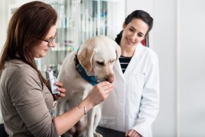
Understanding Veterinary Diagnostics
 In today’s world, if you have a problem with your new car, the diagnostic process is pretty clear. You pull into your mechanic, he hooks your car up to a computer, and within minutes, a complete report is available. Your mechanic changes parts x, y, z and by the end of the visit your car is running like new. When you or your child goes to the doctor, the doctor asks, “Where does it hurt, how are you feeling?” Before you know it, radiographs are ordered, tests performed and a diagnosis is made. Of course I am over simplifying a bit, but in general this is how we expect the diagnostic process to work. In veterinary medicine, this process should run just as smoothly, and in exactly the same way, right? In theory the answer is yes, in practice…well…let me explain.
In veterinary medicine, we start off at a bit of a disadvantage as our patients cannot talk. Although this seems like a funny, simplistic excuse, it truly does come with its own set of problems. Since our patients cannot verbally express to us where they hurt or how they are feeling, we need to learn an entirely different language when dealing with them. An important part of the veterinary diagnostics process is the pet’s history. We also rely strongly on “interpreters,” pet owners and caretakers who provide us with the medical history. In medical terms, a “history” is the signs, symptoms, and collective concerns that lead to the office visit. Understanding the status of the animal prior to the visit helps the veterinarian determine when the signs of illness first began, how severe the signs were, and how constant the signs are. We rely on owners to tell us the pet’s history. Therefore, understanding your pet’s “normal” habits, attitude, appetite, and activity level are of utmost importance.
The Importance of Physical Exam
Of equal importance in the veterinary diagnostic process is the physical examination. This is where the vet clinician truly attempts to listen to the silent, subtle language of illness. Illness has a voice, you just have to be willing to hear it. On a physical examination a pet in pain may subtly offload weight from one limb, they may have a mild limp, they may subtly pull away when a painful area is palpated by the veterinarian. A pet may have a fever, their body may show signs of dehydration, or their eyes may have a dull look. They may be drooling slightly from nausea or turn their head away from food as a wave of queasiness overwhelms them. A pet cannot “tell us in words” where they are lame, sick, or uncomfortable, but they can communicate these things through their actions and behaviors.
After a thorough history and physical examination, hopefully we begin to have some idea why our veterinary patient is not feeling well. Sometimes this means our diagnostic process is complete and a diagnosis with a treatment plan is made, medications are sent home and the pet, hopefully, improves. More often than not, however, our history and physical examination helps us narrow down our list of possible diseases but does not help us definitely diagnose a patient or at least come close enough to put a treatment plan in motion. The phrase “your pet needs further diagnostics” is used frequently but what does it mean?
Labs to Help Diagnose
When we discuss further diagnostics in a general practice setting such as Longwood Veterinary Center, we usually mean bloodwork and lab tests, radiographs, abdominal ultrasound or cytology/biopsy. Blood work includes a Complete Blood Count, or CBC, and a chem screen. A urinalysis is often included as well. In a CBC we are hoping to find normal values for white and red blood cells as well as a normal platelet count. A chem screen evaluates electrolyte balance, kidney, thyroid and liver function. A urinalysis can give us an indication of hydration status, kidney function, and state of infection. There are a multitude of lab tests that can check for very specific diseases if routine blood work (the CBC and chem screen) suggests further tests are needed.
Radiographs and ultrasound are used often in our practice. X-rays help evaluate the bony structures of the body. If a particular joint is painful on our physical examination, we know that joint should be the focus of our radiographic study. X-rays help rule out fractures, infection, bone cancer, and arthritis. Radiographs are sometimes extremely useful in diagnosis of soft tissue related issues as well. For example, a vomiting dog may reveal evidence of dilated intestines and an obvious piece of foreign material in his stomach such as a corn cob, button, or sock. In another example, a middle aged cat with a history of chronic vomiting and diarrhea may have perfectly normal X-rays. An abdominal ultrasound is a better diagnostic step for this kitty as it shows the soft tissue structures of the abdomen in greater detail than radiographs can. Does this mean an abdominal ultrasound can definitely diagnose this kitty’s problem?
 The answer requires a bit of discussion.
An abdominal ultrasound provides us with a tremendous amount of information and can help guide us to an ultimate diagnosis; however, in many situations, the abdominal ultrasound is only the tip of the diagnostic iceberg. Ultrasounds can help us rule in or out multiple conditions and can help us determine the next best course of action. An ultrasonographic evaluation, or ultrasound, as it is frequently called, is a diagnostic tool where ultra high frequency sound waves are passed through a transducer and when directed through the skin can identify structures in the abdomen or other area of interest. Ultrasound examinations are non-invasive and generally well tolerated by most pets, requiring only mild oral sedation to get good images.
Many people are confused when we relay results of an ultrasound only to report a list of possible disease conditions responsible for their pet’s illness but never actually commit to a diagnosis. For example our radiologist may see a tumor in your dog’s liver but we cannot tell you if it is benign or cancerous. In another example, we may tell you your vomiting cat’s stomach and small intestine look thickened on ultrasound, possibly the result of inflammatory bowel disease or lymphoma but we will not commit to a diagnosis of either disease. The ultrasound has allowed us to narrow our list down but for a final diagnosis we may offer to biopsy the abnormal structures. A biopsy is where a piece of tissue is collected from a larger specimen and submitted for microscopic evaluation by a pathologist. A biopsy usually yields a definitive diagnosis.
Unfortunately, most of the time a biopsy requires anesthesia and possibly specialized equipment such as an endoscope to collect. This procedure may not be affordable for many pet owners or the animal may be so ill anesthesia would not be well tolerated. In these cases, the collective results from the physical examination, bloodwork, and ultrasound are taken together to form a treatment plan that is appropriate for the patient and within the owner’s budget. We are basically formulating a “most likely” diagnosis based on all the information we have gathered.
We Care About Your Pets
At Longwood Veterinary Center, the doctors do our very best to thoroughly explain the diagnostic process to you every step of the way. As a pet owner, you must remember that you guide the ship and must feel comfortable with the diagnostic journey. Never hesitate to ask questions or voice any concerns. We are here for you, and are as invested and committed to helping your pet as you are.
