
Heart Disease in Dogs and Cats
February is known for red candy hearts, chocolates, and roses, all expressions of love for our partners, families, and friends. If you are reading this, we know you share your life and heart with a pet, maybe many pets. It seems the perfect time to discuss heart disease in our beloved companions. Heart disease in dogs and cats fall into distinct categories, congenital (pets born with heart disease), disease that is acquired as a pet ages, and disease that occurs as a result of infection or parasitism.
An Introduction to Congenital Heart Disease:
Patent ductus arteriosus (PDA), pulmonic stenosis, ventricular septal defect (VSD), and subaortic stenosis (SAS) are the most common congenital heart defects seen in dogs, thankfully, affecting less than 1% of puppies. The rate of congenital heart disease in kittens is similar to puppies, suspected to affect only 1-2% of kittens. Common defects in kittens include patent ductus arteriosus (PDA), ventricular septal defect (VSD), and mitral valve dysplasia (MVD). Although less common, pulmonic stenosis can also occur in kittens.
Understanding the Most Common Congenital Diseases in Puppies and Kittens:
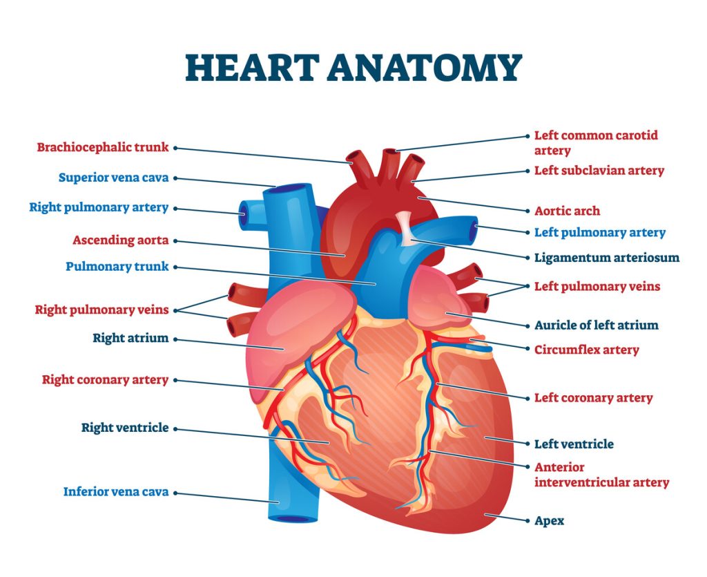
In order to understand heart defects, it is important to first understand normal heart structure. The heart contains four chambers, the right atrium, right ventricle, left atrium, and left ventricle. The four heart chambers are separated internally by septa (thick, muscular walls) and by valves (one-way/directional flaps) that force blood to move in the proper direction and prevent the backflow of blood. By pumping blood throughout the body, the heart helps get rid of carbon dioxide and nitrogen waste while providing the tissues with oxygen-rich blood.
Brief Overview of Common Congenital Conditions:
Patent Ductus Arteriosus (PDA)
All mammals, reptiles, and birds possess a unique structure during embryonic development. A developing embryo does not oxygenate blood via the lungs. Instead, oxygen is transferred to the developing embryo via the placenta (in mammals) or pores in the shell (reptiles and birds).
An embryonic vessel, the ductus arteriosus shunts blood away from the lungs directly to the aorta (to be pumped to the tissues). This small vessel should close within one to three days of birth. This allows all unoxygenated blood to flow from the pulmonary artery to the lungs for oxygenation. In some animals, the vessel does not close. If a PDA is small, the animal often shows no clinical signs of disease. If the PDA is large, excess blood will be forced from the high-pressure aorta into the lower-pressure pulmonary artery. This additional volume of blood then flows to the lungs. The burden of work on the heart increases and results in fluid buildup in the lungs which leads to congestive heart failure. There are many breeds predisposed to this congenital condition including the German Shepherd, Newfoundland, Maltese, Chihuahua, Poodle, Pomeranian, Yorkie, and Doberman (as well as several other breeds). Surgical intervention to close or tie off the aberrant vessel should be performed early in the course of disease to prevent progression to heart failure.
Pulmonic Stenosis
Thickening of the leaflets of the pulmonary valve, the valve between the right ventricle and the pulmonary artery, leads to restriction of the flow of blood going to the lungs. Restriction of flow may be mild, causing little to no clinical signs, or severe (heart murmur, collapse, arrhythmia, exercise intolerance), and possibly heart failure. This congenital anomaly is often coupled with other genetic defects.
Breeds most often affected include brachycephalic breeds, terriers, Samoyeds, and Labradors. However, other breeds such as Boxers and Newfoundlands may also be affected. Treatment may be attempted and may work well for some patients. Using balloon valvuloplasty to enlarge the opening of the pulmonic valve is the treatment of choice.
Ventricular Septal Defect (VSD)
Pets with a VSD literally have a “hole” in their heart. The defect is a hole within the septum, or wall, that separates the ventricles. If small, no clinical signs are seen. If large, blood can be shunted from one side of the heart to the other putting undue strain on that side of the heart. This issue may lead to congestive heart failure. Affected breeds include the English Springer Spaniel, Bulldogs, Westies, and Keeshonds. Surgical closure of the defect requires cardiopulmonary bypass, making it both extremely expensive and not readily available. Other techniques, such as percutaneous transcatheter closure and pulmonary artery banding, may be considered in animals requiring treatment. Luckily, animals with small defects often show minimal clinical signs and do not need intervention. Larger defects, without the resources for surgical intervention, may be managed medically but the prognosis is poor.
Subaortic Stenosis (SAS)
In subaortic stenosis, a scar-like narrowing (stenosis) is present below the aortic valve. This narrowing causes the left ventricle to pump with more effort to move blood through the narrowed opening. This condition is most commonly seen in large breed dogs including the Great Dane, Boxer, German Shepherd, German Short-haired pointer, Dogue de Bordeaux, Golden retriever, Rottweiler, and Newfoundland. Treatment for affected dogs is most commonly limited to medical management. Although open heart surgery to cut open the stenotic tissue or balloon procedures to stretch the narrowing have been attempted, they are not more successful than medical management.
Mitral Valve Dysplasia (MVD)
This congenital disease occurs more commonly in kittens than puppies, especially Ragdolls and Maine Coons. The mitral valve normally keeps blood flowing in one direction from the left atrium to the left ventricle. A malformed mitral valve becomes leaky and blood flows backwards into the left atrium every time the ventricle contracts. This condition may result in congestive heart failure as well as the production of blood clots that can adversely affect other organ systems. Medical therapy for heart failure associated with MVD is the most practical means of treatment although open heart surgery has been used in dogs with the condition.
It goes without saying that dogs with any of these defects should not be used in a breeding program.
The Importance of the Puppy and Kitten Exam
Fortunately, congenital heart disease is not common in companion animals. However, this does not mean evaluating the heart should not play a major role in examination of puppies and kittens. At Longwood Veterinary Center, all young animals receive comprehensive physical examinations at each vaccine visit. This holds true as your pet ages.
Quiet heart murmurs are monitored for any change. The vast majority of benign murmurs will go away by the time young animals reach six months of age. Loud murmurs or murmurs that steadily become louder as a young animal grows are taken very seriously. Our hospital provides the services of a board-certified veterinary Cardiologist and consultations can be scheduled at Longwood Veterinary Center by monthly appointment.
When more urgent evaluation is needed, we are fortunate to live in an area with excellent referral hospitals that have cardiologists on staff. Remember to schedule your puppy or kitten visit today because you love your pets with all your heart and you only want the best for them.
Written by: Corrina Snook Parsons VMD